
Standard cleaning protocols leave measurable Residual Protein in most cannulated instruments. Here’s what three leading hospitals discovered — and what reprocessing teams should do about it.
The reprocessing of reusable medical instruments is one of the most consequential — and most undermonitored — activities in any healthcare facility. While visible soil is easy to address, the invisible threat of residual protein deep inside narrow instrument channels is far harder to detect and far easier to overlook. Residual protein shields microorganisms from disinfectants and sterilants, creating a latent pathway for healthcare-associated infections (HAIs) that standard visual inspection or ATP bioluminescence simply cannot catch.
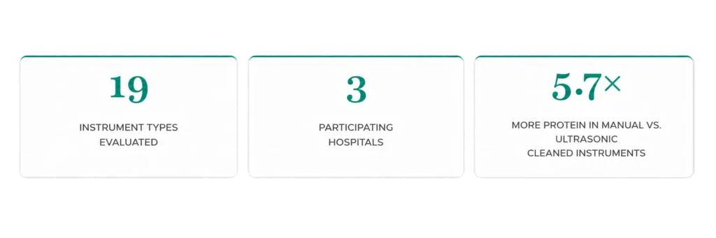
To quantify this risk under real clinical conditions, a multicenter evaluation was conducted across three leading hospitals in Buenos Aires, Argentina: Hospital Italiano de Buenos Aires, Fundación Favaloro (University Hospital), and Hospital El Cruce Dr. Néstor Kirchner. The study assessed 19 distinct cannulated instrument types using the Chemdye® Pro1 Endo quantitative protein detection system — a BCA-based colorimetric assay adapted for internal lumens.
Why Residual Protein in Cannulated Instruments Is a Patient Safety Issue
Cannulated devices — instruments with narrow internal lumens such as ureteroscopes, hysteroscopes, suction tubes, and aspiration cannulas — present unique reprocessing challenges. Their complex geometry limits fluid dynamics, reduces the shear forces needed for soil removal, and creates dead spaces where biofilm and proteinaceous residue can accumulate and persist.
The consequences are well-documented: residual proteins and biofilm impair the efficacy of high-level disinfection (HLD) and steam sterilization. Among cannulated devices, endoscopes represent the greatest cleaning challenge because of their complex geometries, narrow lumens, and diverse material composition. Several outbreaks of multidrug-resistant organisms have been traced directly to inadequate cleaning of these devices, prompting FDA safety communications and accelerated regulatory scrutiny of automated endoscope reprocessors (AERs).
“The persistence of protein residues — even at levels well below visual detection thresholds — constitutes a latent patient safety risk, as these residues can shield microorganisms from disinfectants and sterilants.”
— MULTICENTER STUDY CONCLUSIONS
Guidelines from AAMI ST91 and the CDC support routine, preferably daily, monitoring of manual cleaning processes, and emphasize ongoing staff training and gap analysis. Yet in practice, most reprocessing departments lack the tools to verify cleaning effectiveness at the level of individual instruments and individual lumens.
The Limits of Current Detection Methods
Conventional post-cleaning verication methods each carry signicant limitations when applied to cannulated instruments.
- Visual inspection: cannot detect sub-visible protein Ims or biolm within internal channels
- ATP bioluminescence: not able to detect viruses or prions or even dead cells, which still represent an organic contamination.
- Ninhydrin-based tests and TOC analysis: lack the sensitivity for low-level residues or are impractical for internal lumen access
- Fluorescence imaging: cannot reach or quantify contamination within long narrow, or tortuous lumens.
How the Chemdye® Pro1 Endo System Works
The Chemdye® Pro1 Endo Hygiene Monitoring System is built around the well validated BCA (Bicinchoninic Acid) assay, adapted specically for internal lumen sampling. The system uses Chemdye® SWE high-absorption swabs — available in four diameters (1.7, 2.0, 2.7, and 3.0 mm) and 2.5 meters in length — that traverse the full length of an instrument’s internal channel in a single pass.
DETECTION WORKFLOW
- A size-matched SWE swab is passed through the instrument’s internal channel in a single direction, collecting any residual protein.
- The swab is immersed in the Chemdye® Pro1 Endo reactive solution.
- Proteins reduce Cu²⁺ to Cu⁺, which forms a purple BCA–copper complex proportional to protein concentration.
- The device is incubated at 60 °C for 4 minutes in the Bionova® MiniPro reader.
- Absorbance at 562 nm is measured against a bovine serum albumin (BSA) calibration curve, delivering a quantitative result in μg of protein (range: 1–50 μg; LOD: 0.5 μg).
This design addresses a fundamental limitation of prior methods: no commercially available system could previously access and quantify contamination within long, narrow, or complex lumens. The 2.5 m swab length covers flexible endoscopic channels exceeding 120 cm in a single sampling pass.
Key Findings from the Multicenter Study
1. PROTEIN CONTAMINATION IS WIDESPREAD AFTER STANDARD CLEANING
Across all three hospitals and both cleaning modalities, the Pro1 Endo system detected protein residues in a signicant proportion of instruments that had already completed their institutional reprocessing protocol. Results ranged from 0 μg (undetectable) to 6.3 μg of protein per instrument conrming that standard cleaning does not universally eliminate internal organic contamination.
2. NARROW-LUMEN, LONG-CHANNEL INSTRUMENTS POSE THE GREATEST RISK
When protein results are normalized to internal surface area (μg/cm²), narrow instruments display disproportionately high contamination per unit area. The Olsen Cholangiography Forceps (2 mm diameter, 32 cm channel) showed an average surface density of 0.48 μg/cm², compared to 0.028 μg/cm² for the Ureteroscope and 0.010 μg/cm² for the Cystoscope. Reduced fluid shear, limited mechanical access, and laminar-to-turbulent flow transitions inside narrow channels all contribute to this pattern.

3. Ultrasonic Cleaning Significantly Outperforms Manual Cleaning
Instruments processed via automated ultrasonic cleaning showed an average residual protein level of 0.23 μg, compared to 1.31 μg for manually cleaned instruments — a 5.7-fold difference. Directed cavitation energy accesses complex internal surfaces more reproducibly than manual techniques, which are inherently subject to operator variability.
Critically, however, automated cleaning was not infallible. The Laparoscopic Forceps Sheath (ultrasonic) still registered 1.3 μg in one replicate, and the Cannula Pump showed 1 μg. Instruments with multi-section or multi-lumen architectures may include areas of incomplete cavitation access. Post-cleaning verification remains essential regardless of the cleaning modality.
CLINICAL IMPLICATION
Without post-cleaning verication, instruments carrying residual protein loads
could proceed directly to sterilization — possibly reducing the sterilant penetration and efficacy—
without any opportunity for corrective action.
4. PRO1 ENDO ENABLES REAL-TIME CORRECTIVE ACTION
The most operationally signicant finding of the study was the system’s role as an active feedback tool. At Hospital El Cruce, two instruments with initial readings above 5 μg were agged, re-washed, and retested:
- The Frazier 9 Fr Suction Tube started at >50 μg. Aer one additional manual
wash, the result dropped to 0.5 μg — a greater than 98% reduction. - The Frazier 13 Suction Tube started at 6.3 μg. Two additional manual washes
still le it at 4.9 μg. Only aer switching to ultrasonic cleaning did the result
reach 0 μg.
These cases illustrate the system’s unique value: it does not merely conrm a pass or fail — it identfies when a cleaning method is inadequate for a particular instrument and guides escalation to a more eective approach.
Six Evidence-Based Conclusions for Reprocessing Departments
- Standard cleaning is not universally suficient: Residual protein was found across all three institutions and both cleaning methods, confirming that compliance with FUS alone does not guarantee internal cleanliness in cannulated instruments
- Automate where possible: Ultrasonic cleaning reduces average residual protein by a factor of 57 compared to manual cleaning. Where instrument compatibility permits, automated cleaning should be the preferred modality.
- Prioritize narrow-lumen instruments for enhanced protocols: Instruments with internal diameters s2 mm and longer channels particularly cholangiography forceps, ureteroscopes, and neuro aspiration cannulas-require dedicated monitoring programs.
- Use quantitative, not qualitative, monitoring: Only a quantitative system can guide corrective action and document the degree of improvement aer re-washing.
- Pass/fall Indicators are insucient for complex instruments: Verify every cleaning episode, not just periodic audits.
Inter-procedural variability in soil load and cleaning thoroughness means that a single clean instrument does not predict the next - Document results for QMS and regulatory compliance: Quantitative records from the Bionoval MiniPro reader create a traceable audit trail supporting compliance with 150 15883 AAMI ST79, and accreditation frameworks that require evidence-based reprocessing quality monitoring.
Conclusion
This multicenter study provides clear, real-world evidence that residual protein contamination in reusable cannulated medical instruments is not a theoretical concern — it is a confirmed, measurable phenomenon occurring under standard institutional conditions. The Chemdye® Pro1 Endo system demonstrated superior sensitivity, broad instrument compatibility across all 19 device types evaluated, and direct clinical utility as a real-time corrective tool that no conventional method can replicate.
Adopting quantitative protein monitoring as a routine component of the reprocessing verication workflow represents one of the most actionable steps a facility can take to reduce the risk of device-associated infections in patients undergoing procedures with cannulated instruments.
“Pro1 Endo functions not merely as a passive quality indicator, but as an active component of the cleaning verication loop — one that prevents contaminated instruments from proceeding to sterilization and subsequent patient use.”